PhD students Kadie-Ann Sterling and Clarisse De Vries from the University of Aberdeen iCAIRD team tell us about their research and how artificial intelligence can aid stroke and breast cancer diagnoses.
Come with us to the scene of innovative research and witness history in the making. Discover how a group of scientists at the Translational Healthcare Technologies Group have been able to tinker with UV and fibre optics to create a new cutting-edge piece of technology that will go on to save lives and treat infections and diseases. We have developed this using two things you can find in everyday life: fibre optics and UV light. Fibre optics are used to supply your internet in most areas now. Those same fibres lend themselves to be flexible thin instruments that can explore the body in less invasive ways than open surgical procedures.
UV light is bactericidal (kills bacteria), but it can also be cancer-causing, which is why we cover up or apply sunscreen at the beach. However, we’re planning to use a particular wavelength of UV that means we’ve lost the cancer-causing side of it, meaning what we have is powerful bacteria-killing light rays! We have combined the two to develop this world-changing piece of technology. The fibres, giving us the flexibility to reach places in the human body that are hard and dangerous to reach, such as the lung, and the UV gives us the ability to kill bacteria wherever we find them.
Together we can see this technology used for things that, soon, antibiotics may not be able to be used for due to the increase in super-bacteria’s resistance.
Investigate the part Artificial Intelligence plays in healthcare. Watch Dr Ben Glocker from Imperial College London explain how Artificial Intelligence algorithms are trained to read medical images as seen in the ‘Spot the Lesion’ game.
With thanks to iCAIRD (The Industrial Centre for Artificial Intelligence Research in Digital Diagnostics) for their help with this section.
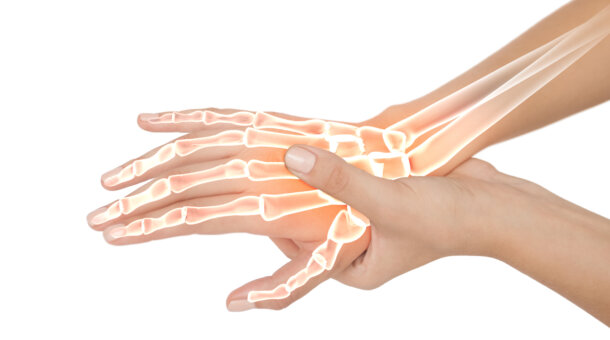
This fantastic resource from the European Space Agency: ESA Kids looks at what’s inside your hand and gives you instructions for building a bionic hand model. Click the link below to download the resource.
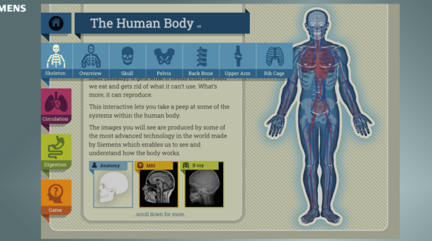
Ultrasound technology is commonly used to support medical diagnosis. These resources developed by Siemens PLC help explain what ultrasound is and investigates the many applications it has. You’ll find a presentation, teaching aids and pupil worksheets designed for 11-14 year olds.
At the level we can see them, medicines look like tablets, pills or liquids. At a much smaller level, tinier than we can see, they have a much wider variety of shapes. They’re often so tiny that we have to use special lab equipment to know what they look like, but each type of medicine has its own shape. They can have rings, lines, all sorts of shapes. They work in 3 dimensions! We can model what they look like. There are modelling kits that you can buy, but it’s also easy enough to do it with things you might find around the house. Fruit can be a surprisingly useful tool for science.
With thanks to the University of Dundee, Wellcome Centre for Anti-Infectives Research (WCAIR) for sharing this resource. For more from WCAIR visit https://wcair.dundee.ac.uk/public-engagement/
Before designing a new medicine it is important that we understand the body, as different treatments need to reach different organs. For example, medicines that treat TB need to target the lungs while medicines that treat malaria need to target the liver and/or the blood.
With thanks to the University of Dundee, Wellcome Centre for Anti-Infectives Research (WCAIR) for sharing this resource. For more from WCAIR visit https://wcair.dundee.ac.uk/public-engagement/
Colon Capsule Endoscopy is a procedure which involves swallowing a capsule the size of a vitamin pill. The capsule contains a digital camera which is swallowed and, on its journey, takes up to 400,000 images (32 per second), which are remotely reviewed and analysed.
It is highly accurate, with the potential to be cost effective, less invasive, and more acceptable to patients, than existing procedures. It could potentially be a viable and safe alternative to traditional colonoscopy. This ambitious project brings together innovation partners from across the NHS, academia and healthcare sectors.
Covid tests have become a part of our daily lives – but what’s the science behind them? Have you ever wondered how tests for viruses like COVID-19 work? In this video, Michelle demonstrates how scientists tests for viruses and explains the difference between a virus test and antibody test. Please note – The science behind vaccines and covid testing is ever changing because of different variants. All information was correct at the time of recording.
Mesothelioma is an unusual type of lung cancer that is extremely difficult to measure and detect. Because of its complex shape it requires highly skilled doctors to identify the tumours and draw them by hand on scans. This leaves room for user error and takes away valuable time that doctors could spend treating patients.
To help solve this problem, experts at the University of Glasgow and Canon Medical Research Europe have created a prototype artificial intelligence (AI) system, which can detect and measure Mesothelioma tumours on CT scans without any human input.
Learn how experts trained the AI system to automatically detect tumours by using existing scans that were marked by doctors. Could this AI tool soon help doctors treat cancer patients with greater precision and for less money? Could it help improve clinical trials to find new drug treatments more quickly?
AI system able to detect and measure complex tumours without any human input
Mesothelioma is a cancer of the lining of the lung caused by inhalation of asbestos, a building material commonly used in construction. Glasgow has the highest incidence of Mesothelioma in the world, due to the historical use of asbestos in its shipbuilding industry.
Asbestos has been banned in the UK since 1999, however we expect to see Mesothelioma cases continue to rise as it can take 30-50 years for the disease to develop after asbestos exposure.
Countries such as Brazil, Russia, India, and China still use asbestos in their construction industries, and so we expect to see global cases rise too.
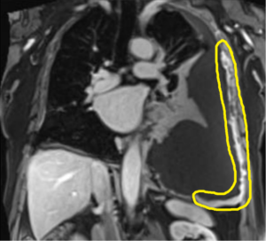
Mesothelioma is unusual because it doesn’t grow like a round ball, like most tumours. Instead, it grows like a ‘rind’ around the surface of the lung, forming a complex shape.
Mesothelioma shown within yellow loop
Think about the orange peel being the tumour rather than a seed in the centre of the orange.
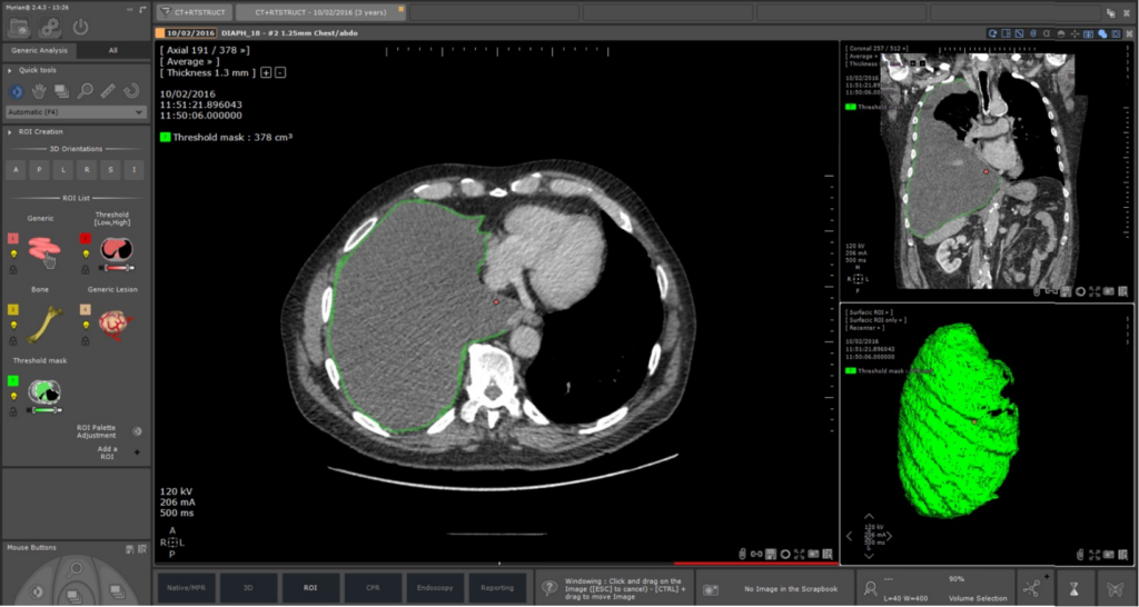
Mesothelioma shown in green
This makes it difficult to detect and measure Mesothelioma on scans and requires highly skilled doctors to identify and quantify the size of the tumour, and any changes with treatment. It involves drawing the tumour on the scans by hand based on their own experience and judgement. This is time consuming, leaves room for user error, and uses up time the doctor could spend treating patients.
Experts at the University of Glasgow and Canon Medical Research Europe (a Scottish firm specialising in next generation medical imaging software) have created a prototype AI system able to automatically find and measure Mesothelioma on CT scans without any human input.
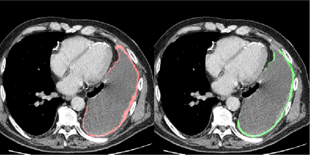
The AI was trained by showing it over 100 CT scans, on which an expert clinician had drawn around all areas of the tumour which showed the AI what to look for. The trained AI was then shown a new set of scans and was able to find and measure the tumour extremely accurately on its own.
Human drawing in red AI drawing in green
The AI tool streamlines tumour measurements, potentially making clinical trials of new drugs more accurate, less time-consuming and therefore less expensive.
This pilot project was funded by Cancer Innovation Challenge, and has been successful in leveraging a multi-million Cancer Research UK Accelerator Award for a Mesothelioma Research Network called PREDICT-Meso.
PREDICT-Meso brings together expert mesothelioma researchers from across Europe to design and validate new therapies and diagnosis techniques for Mesothelioma.
After further work to test and validate the AI tool as part of PREDICT-Meso, the AI technology may soon be available to help doctors measure Mesothelioma on scans during treatment with greater precision and at a reduced cost.
The successful results of the project will provide a strong foundation for similar tools to be developed in the assessment of other cancers.
A recent BBC News article highlights the stories of some mesothelioma patients and how this technology can improve their healthcare journey – https://www.bbc.co.uk/news/uk-scotland-56734407
The way proteins are folded is a rapidly-evolving research area in biology and medicine. However, understanding the 3D structures of these protein molecules and how they relate to diseases such as cancer can be very challenging. To make it easier, researchers at the University of Glasgow have collaborated with Sublime/ Edify to develop software that uses virtual reality technology. Their Molecular Viewer software allows users to view and analyse protein structures in 3D, which can help provide a better understanding of the role of proteins in health and disease.
In this video, Professor Edward Tobias explains the importance of the BRCA1 gene in causing hereditary cancer. Using the Molecular Viewer software, he’ll show you the intricate 3D structure of an important part of the human BRCA1 gene. By looking carefully you’ll see how a tiny change in the DNA can cause a dramatic change in the structure of the BRCA1 protein, which can then cause breast cancer.
Covid-19 has shaped our lives over the past year but what is a virus, how are vaccines developed, and what does it take to be a virologist? Emma Thomson, a professor of infectious diseases at the MRC-University of Glasgow Centre for Virus Research, discusses how scientists have worked together more closely than ever before to implement innovative solutions that have helped to slow the spread of Covid-19. Find out the skills you need to be a successful virologist within a global scientific community that is dedicated to safe guarding public health.
How can artificial intelligence (AI) help doctors?
Artificial intelligence is speeding up the diagnosis of cancer and supporting doctors to make better, faster decisions that could save lives. AI can be much faster than humans at finding tiny differences in images. AI can be trained to look for differences in shape and pattern within similar images.
AI is trained by analysing hundreds of sample images. These images can be of anything, from objects that humans find it easy to recognize, to microscopic things, like cells. The AI looks for patterns in the pixels that it associates with each item. A percentage score represents how certain the AI is about the match.
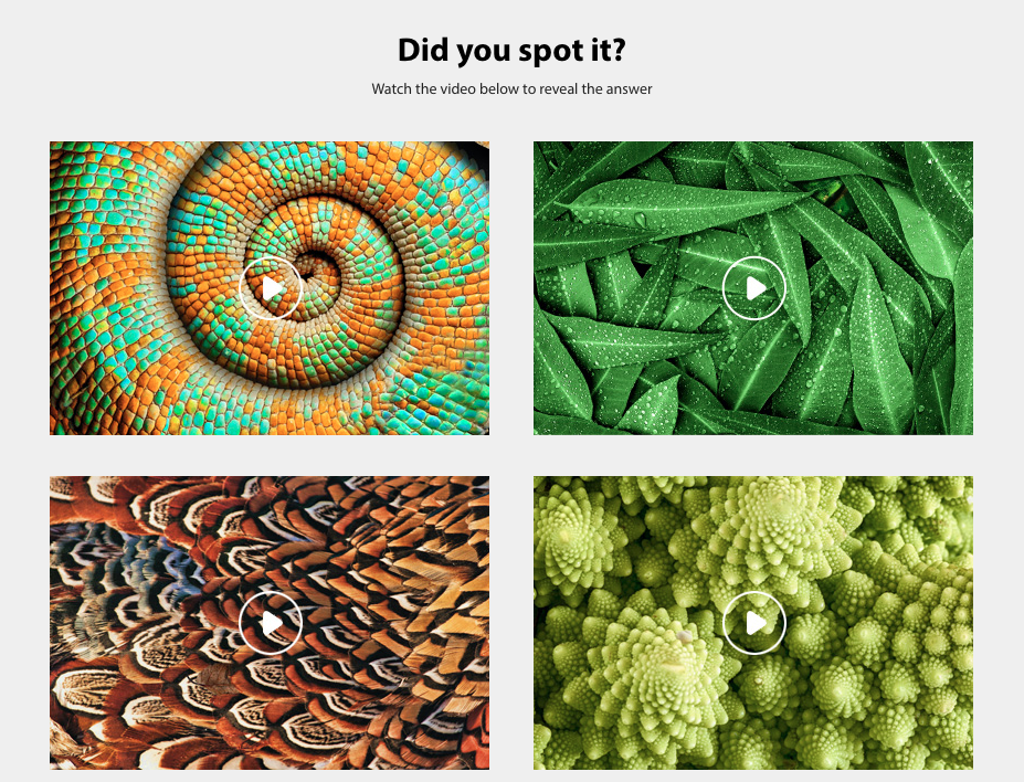
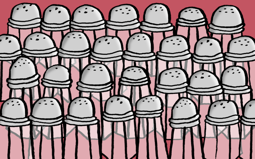
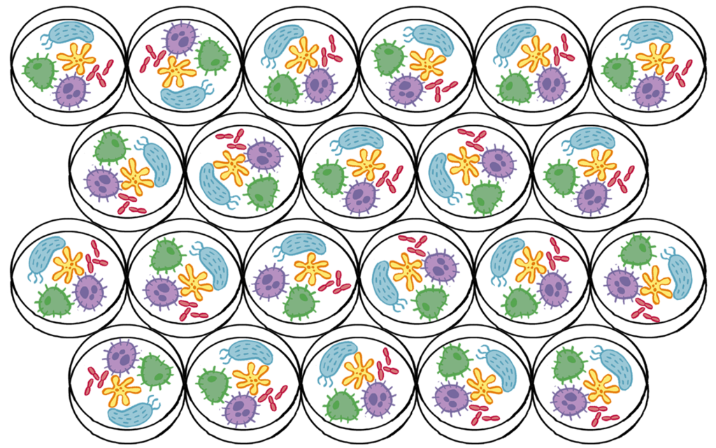
Look through the images below and see if you can spot the odd one out. Answers at the bottom of the page.
Can you spot the pear among the apples?
Can you spot the salt-shaker among the pepper shakers?
Can you spot the snake among the vines?
Can you spot the petri dish that looks different to the rest?
Think you have found the answers?
Looking for the tell-tale differences in shape, colour and size in these pictures is similar to how pathologists search for signs of disease in tissue samples. This process can be very time consuming, but a specially designed AI system can help by quickly spotting even the smallest variations.
Visit us! Try the AI Microscope and find out about machine learning in our Idea No59 Exhibition on Floor 2 of Glasgow Science Centre.
Many medicines that are used today come from the active ingredients found in plants that would have been used in traditional herbal remedies. For example, the well-known painkiller, aspirin comes from willow bark, and artemisinins the malaria treatment, comes from sweet wormwood. Find out more about the garden and medicinal plants in this short video from WCAIR (Wellcome Centre for Anti-Infectives Research).
You can learn even more from the rescource sheets.
Have you ever wondered what’s inside a research lab? Laboratories are cool, fascinating spaces. They are also busy, can smell a bit funky, and are full of chemicals and other hazards. Because of this, it’s tricky for scientists to let many people into their labs in real life. Thankfully, though, Google is here to help.
The Wellcome Centre for Anti-Infectives Research (WCAIR) at the University of Dundee, have created a lab tour on Google Street View, so you can have an explore at your own pace! They’ve added some useful facts to it too, which you should be able to find on the tour. See if you can discover their safety gear, some of their robots, and a few over-achieving members of staff who seem to be working in virtually every lab!
Imagine standing inside a human cell. Not a cell drawn by an artist but a real cell that was scanned by lasers and then sent to a Virtual Reality (VR) headset! Once the data is in the correct format the cells can be 3D printed, incorporated into a CGI-style 3D animation or you could put on a Virtual Reality headset and be transported inside the cell.
The University of Glasgow are using VR technology to potentially radically change the way we teach biology and the way that students learn about the complex 3D structure of biological tissues. See the workflow involved in laser scanning a biological sample and processing it for sophisticated CGI-style animation, fully immersive Virtual Reality and interactive gaming. Looking at biological images is interesting enough but being able walk around the data, and pick it up, takes learning to a whole new level.